• Home
  • About Us
    Our Company Our Vision Our Value
  • Product Center
    Products
    Endoscopy
    Human
    iPS-8000max
    iBS Series
    Veterinary
    VES-100
    Ultrasound
    Mini-Probe EUS
    IM-02M-01
    iMP Series
  • Resource Center
    Videos Documents
  • Media Center
    News Events Blog Client Story
  • Contact Us
    Contact Career
CN
  • Home
  • About Us
    Our Company Our Vision Our Value
  • Product Center

    Products

    Endoscopy
    Human
    iPS-8000max
    iBS Series
    Veterinary
    VES-100
    Ultrasound
    Mini-Probe EUS
    IM-02M-01
    iMP Series
  • Resource Center
    Videos Documents
  • Media Center
    News Events Blog Client Story
  • Contact Us
    Contact Career

Resource Center

Videos
Documents
Home Resource CenterDocumentsClinical Cases

Endoscopy Clinical Case: Esophageal bulge considered tumor

    Author:InnerMed    Browse:2261

▶ The patient is female, 66 years old(From Shenzhen University General Hospital);


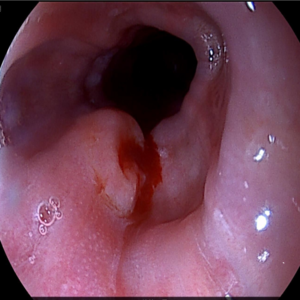
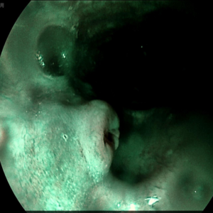
▶ Findings: An umbilicus-like protrusion about 0.8 cm in size was seen on the back wall of esophageal, about 30 cm away from the incisors; the surface of mucosa was congested and edematous; the boundary was clear after NBI observation, the remaining parts of esophageal mucosa was smooth; the vascular network was clear, and no ulcers, erosions, Polyps and varicose veins etc. The cardia was normal. Fundic mucosa was normal. Gastric mucus is clear, and the amount is medium. There was no congestion and edema in gastric body mucosa, and the folds were arranged regularly. The gastric angle is arc-shaped and symmetrical, and the mucosa is smooth. The gastric antrum mucosa is not smooth, red and white, mainly red, and multiple erythema can be seen. The pylorus is round and well opened and closed. There was no deformation, congestion, edema, and ulcer in the duodenal lumen, and a hemispherical protrusion about 0.4 cm in size was seen in the descending segment. The surface mucosa was smooth and normal in color, and the circular folds of mucosa and the mucosa were normal.


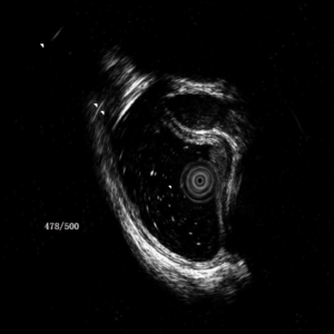
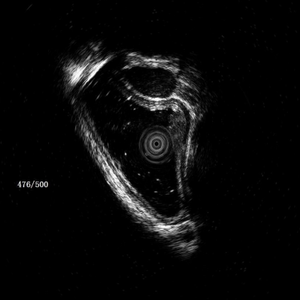
▶ Findings of mini-probe endoscopic ultrasonography: The lesion originates from the mucosa, invades the submucosa and part of the muscularis propria, showing a low mixed echo mass shadow. 2 biopsies were taken.


▶ Diagnostics:

Esophageal bulge considers tumor

chronic non-atrophic gastritis

Consider submucosal cyst in descending part of duodenum protuberance


Share

Previous Article:Mini-Probe EUS Clinical Applications Return Next Article:Endoscopy Clinical Case: Rectal polyps (partial Ca change possible)
Recommended Reading
  • Mini-Probe EUS Clinical Applications

    2023-06-20

  • Endoscopy Clinical Case: Esophageal bulge considered tumor

    2023-06-20

  • Endoscopy Clinical Case: Rectal polyps (partial Ca change possible)

    2023-06-20

  • Endoscopy Clinical Case: Brucella's gland hyperplasia and cyst

    2023-06-20

  • Endoscopy Clinical Case: Colon cancer

    2023-06-20

Follow Us
  • About Us

    Our Company Our Vision Our Value
  • Products

    Endoscopy Ultrasound
  • Resource Center

    Videos Documents
  • Media Center

    News Events Blog Client Story
  • Contact Us

    Contact Career

Hotline

(86-755)26068349

Email

intl-sales@innermed.com

Headquarter

601, Building A2, Chuangzhiyuncheng Project Phase 1, Liuxian Avenue, Xili Street, Nanshan District, Shenzhen, 518000 Guangdong, P.R. China

Links
CE
Privacy Policy | Service Policy | Contact Us
Copyright © 2023 InnerMedical Co., Ltd. All Rights Reserved Designed by Geeke
By continuing your browsing on this site, you agree to the use of cookies in order to optimize the functionality of the innermed.com through the production of anonymous and statistical data. You also agree to the use of third party cookies in order to enable content sharing from and/or to social media provider .